
DMSA Case 5
Details on the Request Card
52 year old female with a heavily calcified, shrunken left kidney with further R upper pole area of calcificationProcedure
Anterior and posterior images are shown, with a right posterior oblique, 3hrs after injection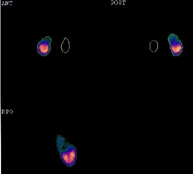
Questions
1) Suggest the most likely diagnosis
2) Would it surprise you if the patient was asymptomatic?
The text is entirely the opinion of the author and does not necessarily reflect that of RUH NHS Trust or the Bristol Radiology Training Scheme. Website content devised by Paul McCoubrie.