
Media Release
Date: 12 September 2019
RUH Radiology Department nears completion
Work has begun to upgrade and replace three scanners at the RUH - the final stage of a development project that will make the hospital's Radiology Department one of the most modern and best-equipped in the South West.
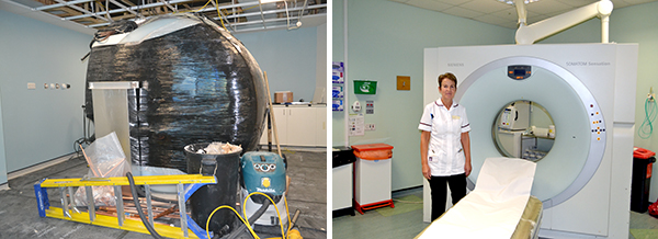
Two older MRI scanners - used to examine most parts of the body, including bones and joints - are being stripped back to their powerful magnet and rebuilt on site with the latest scanning technology. Each scan room that houses them is being refurbished which includes replacing the Radiofrequency cage which lines the room and a complete refit of their control areas.
One CT scanner - which can produce detailed images of the inside of the body, including organs, blood vessels and bones - is being removed and replaced with the latest model.
Its scanning room and control area are also being refurbished.
The work, which is currently taking place and will continue until February 2020, is the final part of a five-year, £7.5m investment programme by the Royal United Hospitals Bath NHS Foundation Trust.
It means the Radiology Department will have six modern MRI and CT scanners as well as X-ray and ultrasound facilities. The department also has a PET-CT scanner used in the detection and diagnosis of cancer and dementia, which was installed in 2016. There are also larger and improved waiting areas for patients.
Di Pressdee, Team Lead Radiographer for CT and MRI said: "This work is the final piece in the jigsaw of our department's major upgrade. It means we will have the very latest in scanning equipment in modern and comfortable surroundings that will enable us to provide the very best services and hospital experience for our patients."
Last year, more than 275,000 patients came for imaging in the Radiology department - of which, over 60,000 were for CT and MRI scans.
ENDS
Notes to Editor
- CT scans can produce detailed images of many structures inside the body, including the internal organs, blood vessels and bones. They can be used to:
- diagnose conditions - including damage to bones, injuries to internal organs, problems with blood flow, strokes and cancer
- guide further tests or treatments - for example, CT scans can help to determine the location, size and shape of a tumour before having radiotherapy, or to allow a doctor to take a needle biopsy (where a small tissue sample is removed using a needle) or drain an abscess
- monitor conditions - including checking the size of tumours during and after cancer treatment
- Magnetic resonance imaging (MRI) is a type of scan that uses strong magnetic fields and radio waves to produce detailed images of the inside of the body.
- An MRI scanner is a large tube that contains powerful magnets. You lie inside the tube during the scan.
- An MRI scan can be used to examine almost any part of the body, including the:
- brain and spinal cord
- bones and joints
- breasts
- heart and blood vessels
- internal organs, such as the liver, womb or prostate gland
- The results of an MRI scan can be used to help diagnose conditions, plan treatments and assess how effective previous treatment has been.
- The RUH Radiology Department undertakes a full range of diagnostic imaging, including PET-CT. The department operates on seven separate sites across the locality undertaking around 293,615 patient examinations per year.