
Respiratory Department
Respiratory Procedures
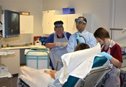
Bronchoscopy
Patients may require a Bronchoscopy to investigate their condition. A Bronchoscopy is an examination of the major lung airways, performed under sedation.
During the procedure a bronchoscope (a fibre-optic flexible tube, the width of a pencil) is inserted through the nose or mouth, and into the major airways. A bronchoscopy is the only test that allows direct viewing of the major lung airways, which may be important in establishing your diagnosis.
During the test, it is possible to obtain a small sample of lung tissue (a biopsy), removed painlessly using tiny forceps passed through the bronchoscope. Additionally it is possible to take a sample washing from your lungs which can be sent for analysis.
You will be required to arrive at the hospital in the morning, and be able to return home at lunchtime. Bronchoscopy is a very safe and well tolerated procedure. If you are due to undergo a Bronchoscopy, you will be provided with more detailed information prior to the procedure.
EBUS-TBNA
EBUS-TBNA (Endo-Bronchial UltraSound-guided Trans-Bronchial Needle Aspiration) is a procedure that allows the doctor to look into your lungs and take samples of the glands in the centre of your chest (mediastinum) using the aid of an ultrasound probe. These glands lie outside the normal breathing tubes (bronchi).
A flexible tube (bronchoscope), which is about the width of a pencil with a bright light on the end is passed into your lungs via your mouth (you will have a mouth guard) with you lying as flat as possible. A small camera at the end of the bronchoscope enables the doctor to look directly into your windpipe (trachea) and breathing tubes (bronchi). A small ultrasound probe on the end of the camera allows the doctor to see the glands in the centre of the chest (mediastinum) and take samples under direct vision. The tissue is removed painlessly through the bronchoscope using a tiny needle. These will be sent for analysis.
EBUS-TBNA is performed as a day case procedure on every Wednesday. You will be asked to attend for the procedure in the morning and will be able to leave hospital at lunchtime. As the procedure is performed under sedation please do not drive yourself and please arrange to be accompanied by family or friends when you arrive and also when you are able to return home.
Thoracosocpy
A thoracoscopy is an examination of the pleural space, which is between the outside of the lungs and the inside of the chest cavity. The doctor will insert a thoracoscope (a tube the width of a pencil with a bright light at the end) between the ribs at the side of the chest wall into the pleural space.
A thoracoscopy is the only test that allows the doctor to view the pleural space directly to assess what may be the cause of the symptoms that you have been experiencing. During this test the doctor will be able to drain off any fluid within the pleural space, carefully examine, and then take a small sample of tissue - a "biopsy". The tissue is removed through the thoracoscope using tiny forceps. This sample will be sent for analysis in the laboratory.
You will be asked to attend the respiratory department in the morning and the thoracoscopy will occur later in the morning. When the examination is finished a chest drain is placed within your chest allowing the lung to re-inflate.
Frequently patients will stay in hospital overnight following the procedure, although on occasions patients may be able to return home later the same day. As the procedure is performed under sedation please do not drive yourself and please arrange to be accompanied by family or friends when you arrive and also when you are able to return home.
Pleurex Catheter
A PleurX catheter is a specially designed small tube inserted in the chest to drain fluid from around your lungs easily whenever it is needed. It avoids the need for repeated injections and chest tubes every time drainage of fluid is needed. The drainage can be performed at home either by a nurse or a relative or friend, whichever suits you. The PleurX catheter is a soft flexible tube that is smaller than a pencil, which remains permanently inside the chest and passes out through the skin. There is a valve on the outer end of the tube to prevent fluid leaking out of the tube.
The pleural space consists of two thin membranes – one lining the lung and the other lining the chest wall. Between these layers, there is a very small space which is usually almost dry. In your case, fluid has collected in this space so that the lung cannot function properly, making you short of breath.
The tube will be put into your chest on the respiratory ward. You will be asked to either sit or lie in a comfortable position by your doctor. You may have an ultrasound examination of your chest prior to the PleurX catheter insertion.
Provided there have been no problems, the catheter insertion is done as a day case and after a short stay at the hospital you will be free to go home a few hours after the procedure. You will be required to have a chest X-ray after the procedure. Someone will need to accompany you home.